


Part 2 of 6 in Circle CVI's Structural Heart series. Every Scan. One Plan. One Platform. A diagnostic cardiac CT report ends in an impression. A structural planning report ends in a set of numbers that a heart team will use to select a device. That is a different product. The impression tolerates hedging, because the clinician reading it brings their own judgment to the question. The planning report does not, because a millimeter of annulus perimeter is not an opinion to be weighed. It is an input to a sizing chart. Imaging physicians have absorbed this shift largely without anyone naming it.

Part 1 of 6 in Circle CVI's Structural Heart series. Every Scan. One Plan. One Platform. The annulus measurement decides the case. Valve choice follows from it. So does sizing, so does the degree of oversizing, so does whether the root tolerates that oversizing, and so does whether you spend the end of an otherwise clean case managing a paravalvular leak. By the time the patient is on the table, that measurement was made days earlier, in a post-processing application the operator may never open, by a reader whose work arrives as a number on a report. For a procedure this dependent on millimeters, that is worth examining.

Part 5 of 5 in Circle's Coronary Plaque series. Also read: Part 1 — How Advanced Plaque Analysis Changes the Clinic al Calculus Part 2 — Th e IT Infrastructure Behind CCTA Plaque Analysis Part 3 — The Financial Case for Coronary Plaque Services Part 4 — D elivering Plaque Analysis Without Disrupting Your Department You have watched the trajectory. Twelve months ago, the conversation about coronary plaque analysis was happening at conferences. Six months ago, it was happening in your referring cardiologists' offices. Now it is happening in your reading room — which lesions are vulnerable, what the total plaque burden is, whether coronary plaque tells a different story than the stenosis grade. That part is good news. Your patients are getting better assessments and the evidence base is catching up to the clinical intuition. On January 1, 2026, the financial case caught up too: the AMA retired the Category III plaque codes (0623T–0626T) and replaced them with a single Category I code, CPT 75577 , for AI-enabled coronary plaque assessment ( ACC Coding Corner ). Plaque analysis is no longer an emerging-technology line item. It is a national fee-schedule procedure. The harder question is operational: is your program set up to deliver it on its own — or to send the studies out and watch most of the reimbursement leave with them? For programs already running CCTA at any meaningful scale, becoming your own plaque lab is more accessible than most assume. It is a workflow choice, not a capital project.
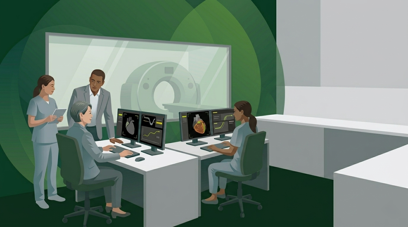
Part 4 of 5 in Circle's Coronary Plaque series. Also read: Part 1 — How Advanced Plaque Analysis Changes the Clinical Calculus Part 2 — The IT Infrastructure Behind CCTA Plaque Analysis Part 3 — The Financial Case for Coronary Plaque Services It's Monday morning review. Throughput is off target again. Two radiologists are working through a backlog of CCTA studies from Friday. Your most experienced cardiac CT tech just submitted a PTO request for a week in July that you can't cover without asking someone else to come in. And now cardiology has sent a note asking why the plaque analysis reports are taking so long. This scenario is not unique to your department. It is the operational reality facing most cardiac imaging programs as CCTA volume grows and clinical expectations evolve faster than workflows do. Coronary plaque analysis has moved from a research capability to a clinical standard — driven by updated ACC/AHA Chest Pain Guidelines , 10-year SCOT-HEART outcomes and the ongoing SCOT-HEART 2 trial , and a growing population of patients and referring physicians who know what to ask for. Meeting that expectation with a manual workflow built for a simpler era of CCTA reporting is not a sustainable operating model. The question is not whether to offer plaque analysis. The question is how to build the workflow to deliver it without adding to a backlog that's already under pressure.