
Part 4 of 5 in Circle's Coronary Plaque series. Also read: Part 1 — How Advanced Plaque Analysis Changes the Clinical Calculus Part 2 — The IT Infrastructure Behind CCTA Plaque Analysis Part 3 — The Financial Case for Coronary Plaque Services It's Monday morning review. Throughput is off target again. Two radiologists are working through a backlog of CCTA studies from Friday. Your most experienced cardiac CT tech just submitted a PTO request for a week in July that you can't cover without asking someone else to come in. And now cardiology has sent a note asking why the plaque analysis reports are taking so long. This scenario is not unique to your department. It is the operational reality facing most cardiac imaging programs as CCTA volume grows and clinical expectations evolve faster than workflows do. Coronary plaque analysis has moved from a research capability to a clinical standard — driven by updated ACC/AHA Chest Pain Guidelines , 10-year SCOT-HEART outcomes and the ongoing SCOT-HEART 2 trial , and a growing population of patients and referring physicians who know what to ask for. Meeting that expectation with a manual workflow built for a simpler era of CCTA reporting is not a sustainable operating model. The question is not whether to offer plaque analysis. The question is how to build the workflow to deliver it without adding to a backlog that's already under pressure.

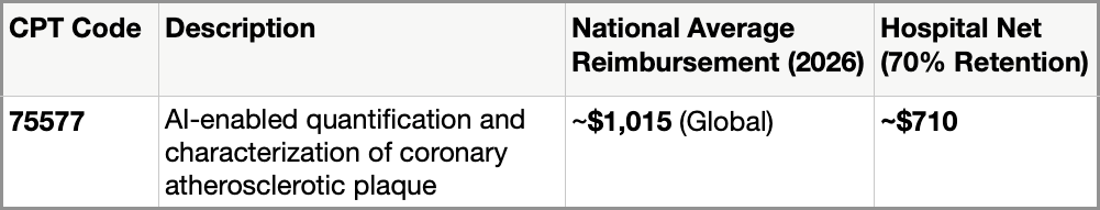
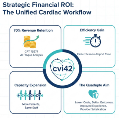
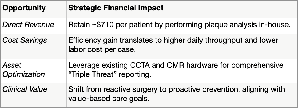
Part 3 of 5 in Circle's Coronary Plaque series. Also read: Part 1 — How Advanced Plaque Analysis Changes the Clinical Calculus Part 2 — The IT Infrastructure Behind CCTA Plaque Analysis The cardiology service line is under familiar financial pressure: rising volumes, tighter margins, growing competition from outpatient and independent imaging centers, and a capital environment that demands every major investment justify itself with a clear return. Against that backdrop, coronary plaque analysis has emerged as a meaningful financial opportunity — one with a growing reimbursement pathway, expanding referral demand, and the kind of clinical differentiation that drives patient retention. But the financial case only materializes if the program is set up to deliver the service efficiently and at scale. This is not an investment in a research capability. It is an investment in a billable, guideline-supported clinical service with a documented and growing payer footprint.
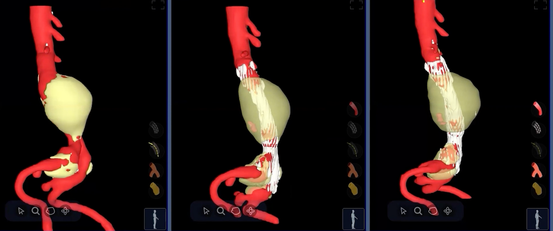
A landmark study shows that measuring how much an aneurysm sac shrinks in the first year after surgery can reliably forecast what that sac's diameter will do over the long haul — unlocking smarter, more personalised patient monitoring. The Problem with Watching Arteries Heal Abdominal aortic aneurysms — dangerous bulges in the body's main artery — kill tens of thousands of people each year when they rupture without warning. Endovascular aneurysm repair, or EVAR , is a minimally invasive surgery in which doctors thread a stent-graft through the groin to seal off the bulge from the inside, like patching a weak hose from within. It's revolutionised vascular surgery, offering patients a far quicker recovery than open surgery. But EVAR is not a cure. The sealed sac still exists inside the body, and over months and years it can change size — ideally shrinking as blood pressure is removed from it, but sometimes stubbornly staying the same or even growing. A sac that keeps expanding after surgery can signal a dangerous leak (called an endoleak ) or graft failure, either of which may require a second intervention. So, after every EVAR procedure, patients face a lifetime of periodic CT scans to check one simple thing: is the sac getting bigger or smaller? "For years, the number clinicians relied on was a single diameter measurement — essentially, how wide is the bulge? But width alone turns out to be a surprisingly blunt instrument." - Background context from the field of post-EVAR surveillance The challenge is that current guidelines require follow-up CT scans roughly every year for life, which is expensive, exposes patients to radiation, and still may miss subtle warning signs until they have become obvious on a simple diameter measurement. Researchers and clinicians have long wondered: is there a better, earlier signal we could use?

Part 2 of 5 in Circle's Coronary Plaque series. Also read: Part 1 — How Advanced Plaque Analysis Changes the Clinical Calculus When clinical cardiology adopts a new capability, IT inherits the infrastructure. And right now, coronary plaque analysis is moving from research tool to clinical standard fast enough that many IT and PACS teams are still catching up. The demand is real. The 2021 ACC/AHA Chest Pain Guidelines made CCTA a Class I recommendation for stable chest pain evaluation. More recent trial data — including 10-year outcomes from SCOT-HEART and the ongoing SCOT-HEART 2 trial — is driving cardiology programs to go further, adding quantitative plaque characterization alongside standard stenosis reporting. That means new software, new data flows, new integrations — and new complexity landing in your environment. How that complexity lands depends almost entirely on the path the department chooses. There are essentially two: a unified platform that performs plaque analysis natively, inside your existing environment — or a send-away service that moves CCTA data out of your network to a vendor cloud, runs the analysis there, and returns a result. Those two paths lead to very different IT outcomes.