Light gray square, with a dark gray border on the sides.

Blog

Seeing the Future of Aortic Repair: Why Volume Matters More Than Diameter

June 9, 2026

A landmark study shows that measuring how much an aneurysm sac shrinks in the first year after surgery can reliably forecast what that sac's diameter will do over the long haul — unlocking smarter, more personalised patient monitoring.


The Problem with Watching Arteries Heal


Abdominal aortic aneurysms — dangerous bulges in the body's main artery — kill tens of thousands of people each year when they rupture without warning. Endovascular aneurysm repair, or EVAR, is a minimally invasive surgery in which doctors thread a stent-graft through the groin to seal off the bulge from the inside, like patching a weak hose from within. It's revolutionised vascular surgery, offering patients a far quicker recovery than open surgery.


But EVAR is not a cure. The sealed sac still exists inside the body, and over months and years it can change size — ideally shrinking as blood pressure is removed from it, but sometimes stubbornly staying the same or even growing. A sac that keeps expanding after surgery can signal a dangerous leak (called an endoleak) or graft failure, either of which may require a second intervention. So, after every EVAR procedure, patients face a lifetime of periodic CT scans to check one simple thing: is the sac getting bigger or smaller?


"For years, the number clinicians relied on was a single diameter measurement — essentially, how wide is the bulge? But width alone turns out to be a surprisingly blunt instrument."

- Background context from the field of post-EVAR surveillance


The challenge is that current guidelines require follow-up CT scans roughly every year for life, which is expensive, exposes patients to radiation, and still may miss subtle warning signs until they have become obvious on a simple diameter measurement. Researchers and clinicians have long wondered: is there a better, earlier signal we could use?

What the New Research Found


A 2026 study published in the Journal of Vascular Surgery tackled exactly this question. The research team looked at patients who underwent infrarenal EVAR — repairs focused on the lower portion of the aorta, just below the kidneys — and tracked two different types of measurements over time: the familiar maximum diameter (width) of the aneurysm sac, and its total volume (how much three-dimensional space it occupied).


Their headline finding was striking: early changes in sac volume, measured in the first months to year after surgery, were reliable predictors of how the sac's diameter would behave over the long term. In other words, if a patient's sac shows meaningful volumetric shrinkage early on, that sac is very likely to continue shrinking in diameter over subsequent years — a good sign. Conversely, a sac that fails to lose volume early tends to show worrying diameter behaviour later.


Why This Matters in Plain English


Think of a partially deflated balloon inside a paper bag. If you measure only how wide it looks from the outside at a single time point, you might miss that it has started to re-inflate slightly in volume. But if you measure how much air — i.e., total volume — is inside at an early stage, you get a far earlier, more complete picture of whether it is truly deflating or not. The study found that volumetric changes early on are a strong forecast of what diameter measurements will show years later.


Why Volume Is a More Sensitive Signal


Diameter measurements have an inherent blind spot. A sac is a three-dimensional object, yet a single diameter captures it in only one direction. It is entirely possible for a sac to expand in volume — accumulating more pressurised blood — while its widest cross-section, known as the maximum sac diameter, barely changes, simply because the growth is happening in a different plane. By the time the width measurement catches up, the problem may already be well established.


Volume, measured by tracing the sac's entire three-dimensional outline on CT imaging, captures this fuller picture. A 5% increase in volume across a three-dimensional structure may represent a clinically meaningful change that a diameter measurement of the same sac would record as essentially stable. Prior research has repeatedly shown that volumetric changes precede diameter changes as a signal of post-EVAR complications — the new study extends this insight by demonstrating that those early volumetric shifts can be used to predict the longer-term diameter trajectory, not just describe the present.

Measurement Type What It Captures Role After EVAR Limitation
Maximum Sac Diameter Widest cross-section of the sac at one plane Standard guideline metric Misses volume changes in other planes; lags behind true sac behaviour
Sac Volume Total 3D space occupied by the sac Early predictive signal Historically requires manual tracing — time-consuming without AI tools

What This Means for Patients and Clinicians


The clinical implications are significant. If early volumetric changes reliably forecast long-term diameter behaviour, clinicians could theoretically use a patient's first post-operative volume measurement to stratify risk — identifying early on who is on a safe trajectory toward sac shrinkage and who may need closer monitoring or earlier re-intervention.


This could mean that patients with strongly regressing sacs could safely have their surveillance intervals extended — fewer CT scans, less radiation, lower cost. Meanwhile, patients showing early volumetric warning signs could be flagged for intensified follow-up before any diameter-based guideline threshold is ever crossed.


Current Society for Vascular Surgery guidelines still define significant sac change as a 5 mm or greater shift in diameter. The new research suggests this binary threshold, while useful, leaves important predictive information on the table. 


Volume change early in the post-operative period is not just a companion metric — it is an upstream predictor of the very metric everyone is already watching.


"The ability to forecast long-term sac behaviour from early volumetric measurements would allow us to move from reactive surveillance to truly proactive, personalised follow-up care."

- Concept from post-EVAR surveillance research


The Practical Barrier — And How AI Is Removing It


Despite volumetric measurement's advantages, it has historically been underused in routine clinical practice. The reason is simple: manually tracing the boundaries of an aneurysm sac across dozens of CT image slices is painstaking, time-consuming work that can add 20 minutes or more to a radiologist's workflow per patient. In a busy hospital, that adds up fast.


This is precisely where artificial intelligence changes the equation. Modern AI-powered segmentation algorithms can automatically identify and trace the aneurysm sac across a full CT scan in under three minutes — with accuracy comparable to manually corrected expert segmentation. The bottleneck that once made volumetrics impractical dissolves.


  • Speed — from 22 min to under 3
  • AI segmentation reduces per-patient measurement time by over 85%, making volumetrics viable at scale.


  • Accuracy on par with experts
  • Studies show AI volumetry closely matches manually corrected senior surgeon segmentation across thousands of slices.


  • Consistent, reproducible results
  • AI removes inter-observer variability that plagues manual measurement, especially across different imaging centres.


  • Better risk stratification
  • Consistent volumetric data over time enables AI-driven classification of sac evolution, improving surveillance decisions.


cvi42 | Vascular CT


This is exactly the kind of clinical advance that cvi42 | Vascular CT is designed to enable. In March 2026, Circle Cardiovascular Imaging expanded its flagship cvi42 platform by integrating AI-driven vascular analysis technology developed by Astute Imaging — bringing automated, quantitative vascular CT analysis into the same unified platform already trusted for cardiac imaging by over 2,100 hospitals across 90+ countries.


The platform supports the full continuum of EVAR care: pre-operative anatomical planning, intra-operative device sizing via AI-enabled virtual device simulation, and — critically — post-operative longitudinal surveillance including automated aneurysm sac segmentation and volumetric tracking. For every follow-up CT scan, cvi42 can automatically generate volumetric measurements, flag changes relative to prior studies, and provide the kind of 3D sac evolution data that the new JVS study identifies as a key predictor of long-term outcomes.


The platform supports the full continuum of EVAR care: pre-operative anatomical planning, intra-operative device sizing via AI-enabled virtual device simulation, and — critically — post-operative longitudinal surveillance including automated aneurysm sac segmentation and volumetric tracking. For every follow-up CT scan, cvi42 can automatically generate volumetric measurements, flag changes relative to prior studies, and provide the kind of 3D sac evolution data that the new JVS study identifies as a key predictor of long-term outcomes.


The result is a workflow where the early predictive volumetric signal described in this research can be captured routinely, rapidly, and reproducibly — without adding burden to an already stretched radiology team.


  • Automated Segmentation – aorta, thrombus, lumen, dissection – fully AI driven
  • Volumetric Tracking – Longitudinal sac volume change over serial studies
  • Virtual device planning – AI-enabled stent-graft sizing and anatomical fit simulation
  • Unified cardiac and vascular workflow – one platform for all CV imaging needs


The Bigger Picture: From Surveillance to Prediction


The shift from measuring what has happened to predicting what will happen is one of the defining ambitions of modern imaging. Diameter tells you where a sac is today. Volume — especially early volume change — tells you where it is headed.


As the evidence base grows, volumetric thresholds may begin to inform surveillance guidelines the way diameter thresholds do today. Hospitals that already have automated volumetric tools in place will be well positioned to act on these evolving recommendations — and to participate in the research studies that will shape them.


More immediately, the findings from this study offer a compelling argument for any vascular or radiology programme still relying solely on diameter: you may be watching the right thing but starting too late. The signal that predicts where a sac is going is available from the very first post-operative scan, in three dimensions, if you have the tools to read it.


The research discussed in this post reinforces a growing body of evidence that volumetric sac analysis after EVAR delivers clinical intelligence that diameter measurement alone cannot provide — and that AI-enabled platforms like cvi42 | Vascular CT are making that intelligence practical at the point of care. 


Primary source: "Early Sac Volume Changes Predict Long-term Diameter Sac Dynamics After Infrarenal EVAR." Journal of Vascular Surgery, 2026. View original paper →

June 2, 2026
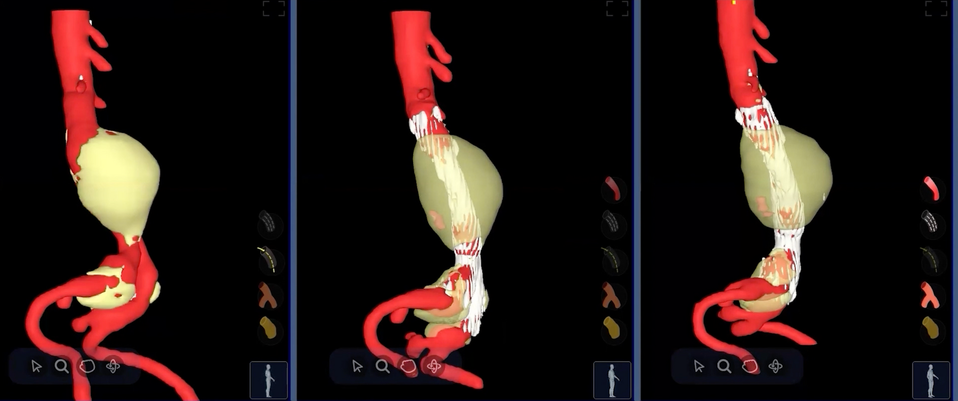
Part 2 of 5 in Circle's Coronary Plaque series. Also read: Part 1 — How Advanced Plaque Analysis Changes the Clinical Calculus When clinical cardiology adopts a new capability, IT inherits the infrastructure. And right now, coronary plaque analysis is moving from research tool to clinical standard fast enough that many IT and PACS teams are still catching up. The demand is real. The 2021 ACC/AHA Chest Pain Guidelines made CCTA a Class I recommendation for stable chest pain evaluation. More recent trial data — including 10-year outcomes from SCOT-HEART and the ongoing SCOT-HEART 2 trial — is driving cardiology programs to go further, adding quantitative plaque characterization alongside standard stenosis reporting. That means new software, new data flows, new integrations — and new complexity landing in your environment. How that complexity lands depends almost entirely on the path the department chooses. There are essentially two: a unified platform that performs plaque analysis natively, inside your existing environment — or a send-away service that moves CCTA data out of your network to a vendor cloud, runs the analysis there, and returns a result. Those two paths lead to very different IT outcomes.
Two people talk across a desk in a green-toned office, with a coronary CTA in the background.
May 19, 2026
Part 1 of 5 in Circle's Coronary Plaque series. Consider a familiar scenario. A 52-year-old presents with atypical chest discomfort. The stress test is borderline. The referring physician is uncertain — is this true coronary disease, or something else? Standard imaging has been done. The anatomy looks, on the surface, mostly normal. And yet something about this patient doesn't sit right. This is the case where clinicians have historically had to make consequential decisions with incomplete information. Escalate to invasive angiography and risk an unnecessary procedure. De-escalate and risk missing a vulnerable plaque that hasn't yet caused significant stenosis — but will. Coronary artery disease doesn't announce itself neatly. A substantial share of acute coronary events occur in patients with non-obstructive coronary disease — lesions that would not have triggered revascularization on a standard angiogram. The stenosis grade has always been an imperfect surrogate for risk. What matters is the plaque itself: its composition, its burden, and its vulnerability. Advanced CCTA plaque analysis changes that calculus.
A medical professional in a navy blue scrub top points to heart scan imagery on a monitor while consulting a colleague.
May 12, 2026
The Effort, Risks, and Why It’s Worth It Quantifying the Effort of a Single Platform Learning a new (or deeper) way of working Even for existing cvi42 users, full standardization across cardiac imaging means: Adapting to new modules or modalities. Rethinking how you structure reading lists when more work is visible in one place. Helping develop shared templates and protocols. It’s completely normal to see a temporary dip in productivity when something new is introduced. Our brains are wired for loss aversion: we tend to focus more on what we might lose than on what we could gain. Recognizing that bias helps put the slowdown in perspective: it’s not a setback, it’s just part of the process that leads to long‑term progress.
Secure data within the cvi42 cardiovascular imaging application for AI enabled postprocessing
May 5, 2026
Local Processing for Complete Data Security When cvi 42 processes imaging data, everything takes place within the customer’s secure environment. All image data and derived results are managed locally, whether on a hospital workstation or through a customer-managed server installation. No data is ever transmitted outside the institution. This architecture ensures compliance with strict hospital IT policies and data protection frameworks. For clinical users, this means AI-powered results without any compromise to data privacy or network security. The Circle AI Engine: Trained, Validated, and Frozen “Each of the AI models powering cvi42 is architected and developed within Circle’s controlled research and development environment. Circle’s data science and clinical AI research teams use diverse and representational datasets to train and validate each algorithm. The process typically involves supervised learning, where the AI learns to recognize patterns and structures such as the left ventricle, myocardium, or aortic root by comparing its results to expert-annotated data. Once performance meets clinical and regulatory standards, the AI model is locked, “frozen” and encrypted during its integration within cvi42. This means the model’s behavior is fixed, it does not continue to learn or change once deployed at a customer site. The model you use in cvi 42 is the validated version approved for clinical use, ensuring consistent and reproducible results across all installations. No Learning from Customer Data It is important to clarify: the AI in cvi 42 does not learn from any data processed at the customer site. The algorithm applies its pre-trained parameters to each image set locally. It does not store patient data, send information externally, or modify its internal model based on what it sees or whether a user edits its outputs. Each analysis is isolated, ensuring the AI’s decisions remain consistent and the patient’s information stays protected within the facility’s network. How the AI Analyzes Medical Images At a technical level, cvi42’s AI is a deep learning-based image analysis engine trained to recognize and segment cardiac anatomy on MR and CT images. Primarily using convolutional neural networks, it performs pixel- or voxel-level classification to delineate key structures, including the endocardial and epicardial borders. These segmentations enable the measurements of clinically relevant metrics such as chamber volumes, ejection fraction, and myocardial mass. This process mimics how expert readers would interpret the same dataset, but it happens in seconds and with objective consistency across cases. Designed for Trust, Built for Performance AI in cvi 42 is designed to automate routine analysis while keeping clinicians fully in control. Users can review, adjust, and approve AI-generated contours as needed, ensuring that results always meet their clinical standards. Combined with local data processing, frozen AI models, and Circle’s rigorous training pipeline, this approach delivers accuracy and reliability without ever compromising patient privacy.

Subscribe to our newsletter

 Don’t miss future articles or publications.