CHICAGO and CALGARY, Alberta – (Oct. 22, 2026) – GE HealthCare (Nasdaq: GEHC) and Circle Cardiovascular Imaging Inc. (Circle CVI) today announced the introduction of a coordinated quantitative cardiac magnetic resonance (CMR) perfusion workflow combining
MyoFlow, GE HealthCare’s dual-sequence cardiac stress perfusion acquisition, with
cvi42 | Quantitative Perfusion, Circle CVI’s automated quantitative analysis software.
Cardiovascular disease remains a significant global health burden, with an estimated 254 million people living with ischemic heart disease in 2021 . Cardiac MR can provide a comprehensive view of cardiac anatomy, function and tissue, but advanced CMR exams can be technically demanding and remain a relatively small part of overall MR utilization. MyoFlow and cvi42 are designed to help address some of these workflow challenges by simplifying stress perfusion acquisition and automating quantitative analysis.
GE HealthCare is bringing together devices and digital capabilities to help clinicians access the right insights at the right time across each patient’s care journey. Stress cardiac MRI, a guideline-recommended test for myocardial ischemia, provides a comprehensive view of heart perfusion, function, and scar tissue in a single exam. Together, MyoFlow and cvi42 simplify image acquisition and automate analysis, delivering patient-specific measurements that support diagnosis and treatment decisions.
“Advanced cardiac MR is most valuable when clinicians can move efficiently from image acquisition to information they can interpret and act on,” said Fotis Vlachos, Chief Marketing Officer, MR and Women’s Health at GE HealthCare. “By bringing together a simplified GE HealthCare acquisition approach with Circle CVI’s quantitative analysis, we are working toward a more connected cardiac MR experience that keeps clinicians at the center of interpretation and decision-making.”
The collaboration expands GE HealthCare’s disease-focused cardiac MR portfolio and Circle CVI’s role in advanced cardiovascular analysis. For GE HealthCare cardiac MR customers, it creates a coordinated path from acquisition through quantitative analysis, visualization and reporting, while keeping clinicians in control of review, interpretation and clinical decision-making.
- MyoFlow dual sequence is available on GE HealthCare SIGNA MRI scanners running MR31 or later software. Not available in all markets.
- cvi42 is CE marked and available for sale in EU member states. cvi42 | Quantitative Perfusion is available for research use only in the United States. MyoFlow and cvi42 | Quantitative Perfusion are separate offerings supplied by GE HealthCare and Circle CVI, respectively. MyoFlow supports cardiac stress perfusion acquisition; quantitative analysis is performed in cvi42 and requires the applicable Circle CVI software license. The coordinated workflow will initially be offered in selected European markets, subject to country-specific regulatory and commercial availability. Product configuration, indications and availability vary by country. Contrast agent use and pharmacologic stress should follow applicable product labeling, institutional protocols and clinical guidelines.
- Yang, L., Zheng, B., & Gong, Y. (2025). Global, regional and national burden of ischemic heart disease and its attributable risk factors from 1990 to 2021: A systematic analysis of the Global Burden of Disease Study 2021. BMC Cardiovascular Disorders, 25, 625.
From a complex perfusion exam to a streamlined quantitative workflow
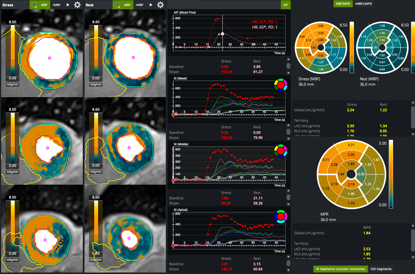
Designed to make advanced stress perfusion more practical, MyoFlow supports each first-pass perfusion scan as one continuous, free-breathing acquisition with a single contrast bolus. The dual-sequence technique captures the arterial input function and myocardial perfusion images within the same acquisition. Circle CVI’s cvi42 then automatically generates pixel-wise maps and American Heart Association (AHA) 16-segment polar maps of stress and rest Myocardial Blood Flow (MBF) and Myocardial Perfusion Reserve (MPR).
Stress perfusion CMR evaluates how well blood reaches the heart muscle during pharmacologic stress. Visual interpretation remains central to clinical practice, but relative contrast differences can be more difficult to assess when perfusion reductions are diffused or involve multiple coronary territories.
MyoFlow is designed to simplify acquisition by collecting arterial input function and myocardial perfusion data during the same first-pass scan. Free-breathing acquisition can reduce dependence on breath-hold performance, while a single bolus per first-pass acquisition avoids the additional complexity of a separate pre-bolus. In cvi42, automated contour detection and polar map analysis reduce manual post-processing steps, and produces reliable, reproducible measurements even under challenging imaging conditions (e.g., motion, noise, surface coil intensity bias). Together, these capabilities are intended to promote more consistent workflows, support diagnostic confidence and help lower the training burden for readers using advanced perfusion assessment.
“Quantitative perfusion adds objective myocardial blood flow and perfusion reserve measurements to visual assessment and can help characterize both regional and diffuse perfusion abnormalities,” said Kevin Steel, M.D., Chief Medical Officer, Circle Cardiovascular Imaging. “Pairing GE HealthCare's dual-sequence acquisition with Circle CVI's automated pixel-wise analysis is designed to make advanced perfusion assessment more practical, reproducible and accessible for cardiac MR teams.”
GE HealthCare Media Contact:
Katie Scrivano
M +1 262-512-5281
Katherine.Scrivano@gehealthcare.com
Circle Cardiovascular Imaging Media Contact:
Circle Cardiovascular Imaging Inc.
marketing@circlecvi.com | circlecvi.com
About GE HealthCare Technologies Inc.
GE HealthCare is a leading global healthcare solutions provider of advanced medical technology, pharmaceutical diagnostics, and AI, cloud and software solutions that help clinicians tackle the world’s most complex diseases. Serving patients and providers for 130 years, GE HealthCare is delivering bold innovations designed for the next era of medicine across its Advanced Imaging Solutions, Pharmaceutical Diagnostics and Patient Care Solutions segments to help clinicians deliver more personalized, precise patient care. We are a $20.6 billion business with approximately 54,000 colleagues working to create a world where healthcare has no limits.
GE HealthCare is proud to be among
2026 Fortune World’s Most Admired Companies™.