The Future of Cardiac Imaging
CT and MR in Action
Minimised manual adjustment of automated analysis outputs
Introduction and Background
Dr Elisa Rauseo is a Consultant Cardiologist working at Barts Health NHS Trust (London), and a Clinical Senior
Lecturer at Queen Mary University of London. Her expertise focuses on the integration of multi-modality imaging
and artificial intelligence from both clinical and research perspectives. Dr Rauseo explains how cvi42’s automated
segmentation supports cardiac MRI reporting and explores multi-modality cardiac CT/MR workflows.
A Researcher’s Perspective
Drawing on her research experience, Dr Rauseo discusses
the potential benefits of image analysis platforms capable of
handling both MR and CT data:
“From a personal standpoint, dual modality, having both CT and MR together on the same platform represents a great opportunity for research-driven environments and for future methodological developments. In the absence of this, the need is to work across different software tools can inherently lead to delays in the outcome of the analysis.”

Circle’s cvi42 in Action for Cardiac MRI
Consistency of measurements over time is of utmost importance for clinical interpretation and follow-up, this is where cvi42 has been seen to demonstrate a dramatically reduced need for manual segmentation.
“I’d say a reduction of around 50-60% of cases needing to be revisited in some capacity today with the software’ explains Dr Rauseo. The structured and reproducible analysis provided by cvi42 is of particular interest in certain conditions which, for example, would require longitudinal analysis. These would include cardiomyopathies, and heart failure where consistency in measurements over time is important for clinical interpretation and follow up.
Multimodality Informs Clinical Interpretation
In the her field of research, Dr Rauseo has found that the use of multiple imaging modalities has provided complementary insights into cardiac structure and pathology. For example, cardiac MRI with late gadolinium enhancement allows detailed assessment of myocardial tissue characteristics, while cardiac CT, typically performed for coronary evaluation, can, under specific conditions, offer additional information on myocardial enhancement patterns.
Correlating findings across MR and CT may help generate hypotheses around disease mechanisms and support more nuanced interpretation, particularly in complex cases.
Clinical Precision with User Control
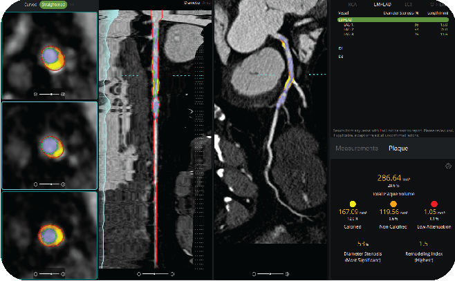
The semi-automated coronary lumen segmentation and plaque quantification
tools provide detailed per-vessel and global measurements, including:
- Calcified plaque volume and burden
- Non-calcified plaque volume
- Low-attenuation, or vulnerable, plaque assessment
All measurements remain editable and user adjustable. Clinicians maintain full oversight of the final report while benefiting from AI-supported software. The result is a reproducible approach that improves consistency between studies and clinicians, while also strengthening longitudinal tracking of disease progression.
Seamless Implementation
Implementation was straightforward. Circle CVI’s technical team provided responsive support for the IT integration,
including PACS connectivity and user onboarding.
According to Dr. Cheong, “The implementation is simple.” The transition required minimal disruption, allowing the
team to quickly realize both clinical and operational benefits.
Technology that Transforms Workflows
The impact extends beyond plaque quantification. Advanced visualization tools have significantly accelerated image processing. Tasks that once required repeated manual adjustments are now automated and can be completed almost instantly, improving efficiency without compromising accuracy.
“The VR and cMPR are on steroids — in the past, it took 1 minute to get a CMPR for each vessel. Now it’s literally 3 seconds. So cool!”
Dr. Benjamin Cheong
A Meaningful Step Forward in Coronary Imaging
For Live Healthy Imaging, adopting cvi42 | Plaque represents more than a workflow improvement. It supports their broader mission to deliver comprehensive, high-quality coronary imaging care. By combining reproducible AI-enabled quantification with in-house control and dramatically reduced turnaround times, the center has established a higher standard for patient-centered cardiovascular imaging.
Dr. Cheong summarizes:
“The cvi42 | Plaque module is an important step in our goal of providing comprehensive coronary imaging care. It substantially reduces the time required for result and report generation while delivering reproducible, AI-enabled coronary plaque quantification locally in a system that is easy to implement in our practice.”
Live Healthy Imaging continues to rely on Circle CVI’s evolving technology to support clinical excellence and continuously improve the delivery of patient care.